Combined linear and non-linear optical imaging of the extracellular matrix
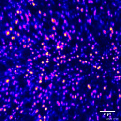
The Extra Cellular Matrix (ECM) provides structural and biomechanical support to cellular constituents in organs and tissue, with variation in composition and function depending on its environment. The ultrastructure of the ECM consists in part of proteins (collagen and elastin for example) whose organization can be observed from the nanoscopic to the macroscopic level through various optical techniques. Understanding the ECM functional role in biological environments may be used to pinpoint early pathological events and consequently devise new treatment paradigms. To translate this knowledge into clinical methods, a new understanding of nanoscopic to macroscopic light-tissue interaction needs to be achieved. By utilizing non-linear microscopic techniques such as Second Harmonic Generation and Adaptive Optics we obtain extreme high-resolution structural assessment of the living ECM. We utilize this data as a framework for our computational models of light transfer to ultimately refine our understanding of the complex interaction between light and biological matter. Based on this knowledge we ultimately aim at improving more translationable optical techniques, such as Mueller Matrix polarimetry, and create quantitative diagnostic tools for the clinical setting. In this context, we are interested in understanding the role of cervical collagen during parturition and determine if early signs of preterm labor can be observed through Mueller Matrix polarimetry, we are also interested in studying the ECM organization in the human cornea and heart valves to guide early intervention.
More about our approach in these publications
- Xuan Liu, Jessica C. Ramella-Roman, Jin Kang, “Robust spectral-domain optical coherence tomography speckle model and its cross-correlation coefficient analysis,” JOSA A 30, 1, 51–59 (2013)
- Xuan Liu, Yong Huang, Jessica C. Ramella-Roman, Scott A. Mathews, and Jin U. Kang, “Quantitative transverse flow measurement using optical coherence tomography speckle decorrelation analysis,” Opt. Lett. 38, 805-807 (2013)
- L. Luu, P. A. Roman, Scott A. Mathews, J.C. Ramella-Roman, “Microfluidics based phantoms of superficial vascular network,” Biomedical Optics Express, 3(6), 1350-1364, (2012)
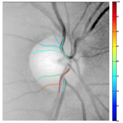
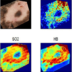
- P. Lemaillet and J.C. Ramella-Roman, “An eye phantom for measurement of retinal oxygenation,” J. of Biomedical Optics, 14, 064008, (2009)
- J.C. Ramella-Roman, S.A. Mathews, H. Kandimalla, A. Nabili, D.D. Duncan, S.A. D’Anna, S.M. Shah, Q.Q. Nguyen, “Measurement of oxygen saturation in the retina with a spectroscopic sensitive multi aperture camera,” Optics Express, 16, 6170-6182, (2008)
Support
We gratefully acknowledge the support of the following agencies during this research effort:- The Herbert Wertheim Foundation
- STROBE: A National Science Foundation Science and Technology Center under Grant No. DMR 1548924.