Background
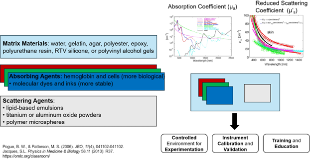
Tissue-mimicking phantoms are fabricated and used to evaluate the performance of wearable devices and SpO2 sensors and investigate potential sources of error. These phantoms offer a low-cost, well-controlled solution, eliminating the need for expensive and time-consuming in-vivo trials. A general overwiew is present below:

Molded
We are able to mold optical phantoms using materials like silicone,
incorporating scattering and absorbing agents to replicate the optical properties of biological tissues for various
imaging and monitoring applications. To achieve this, we may use absorbers like carbon black, India Ink, and Silicon Pigment dye,
creating concentration curves by dissolving these agents in water and IPA, respectively, and measuring their absorbance at wavelengths of interest.
These curves help us prepare specific concentrated solutions to accurately simulate tissue layers and blood.
Available materials: Silicone A-B, Sylgard-184 PDMS, Polyurethane.
3D Printing
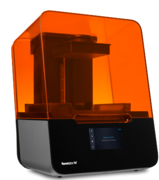
We use 3D Printing to create optical phantoms by precisely fabricating complex geometries and embedding scattering and absorbing agents. This approach offers advantages such as customization, reproducibility, and rapid prototyping. Our 3D-printed phantoms are used to assess and adjust the optical properties, including those related to blood simulation, by integrating BMFs and evaluating their effects.
Available materials: Clear V4, Elastic 50A, Silicone 40A, PLA, ABS, PVA, TPU 85A, TPE 95A.

Dynamic Phantoms
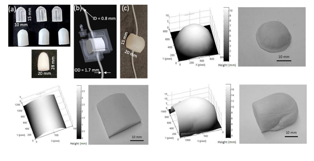
We develop phantoms that mimic arterial pulsation to meet the needs of optical methodologies like Photoplethysmography (PPG), Pulse Oximetry, and specific ophthalmology systems. We cast and mold wrist tissue phantoms to replicate actual tissues' optical and mechanical properties. We control vessel diamter and depth in mold design and adjust it to represent different Body Mass Indexes (BMIs). We utilize a closed-loop Test Bed to obtain PPG data by pumping blood-mimicking fluids inside our phantoms. This test bed allows easy use and adapts to any wrist- and finger-worn wearable device. An integrating sphere characterizes the optical properties of these phantoms using an adding-doubling method.
Other Phantoms

Interested? How to get involved!
Contact: Ajmal (ajmal003@fiu.edu) or Andres (andrrodr@fiu.edu) for more info on our phantoms.